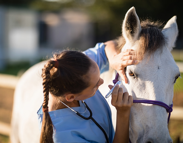
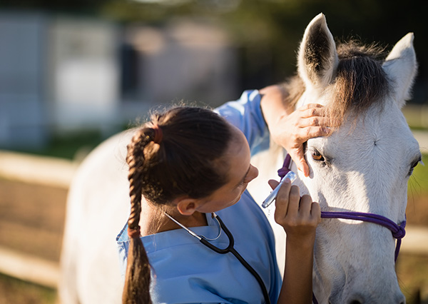
WHAT IT TAKES TO DIAGNOSE PPID
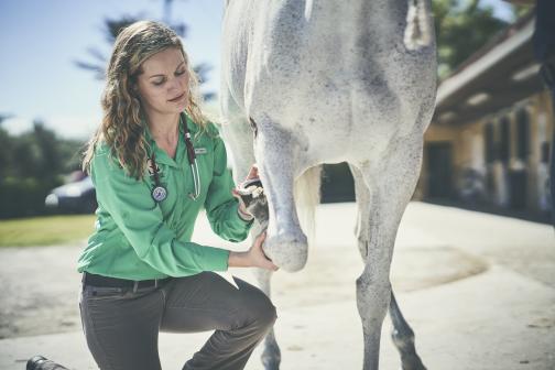
PPID may present through a collection of subtle clinical signs. To accurately diagnose, use a combination of the following:
- Horse's history
- Complete physical examination
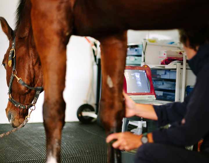
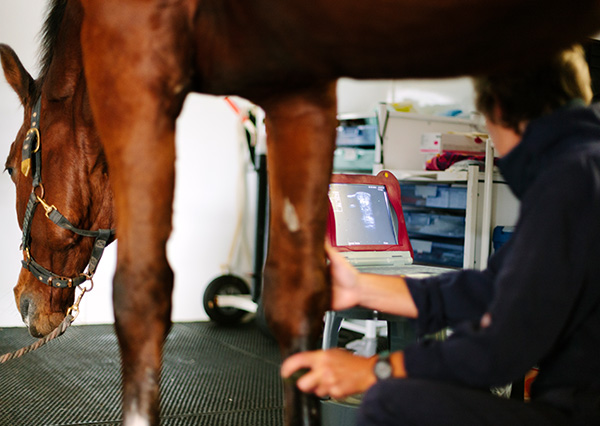
- Appropriate diagnostic evaluation
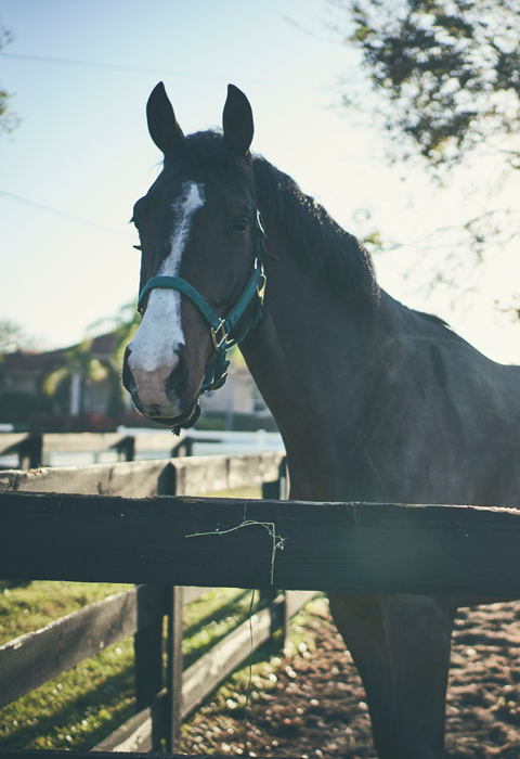
PPID DOESN'T DISCRIMINATE
PPID was once thought of as an old horse disease.
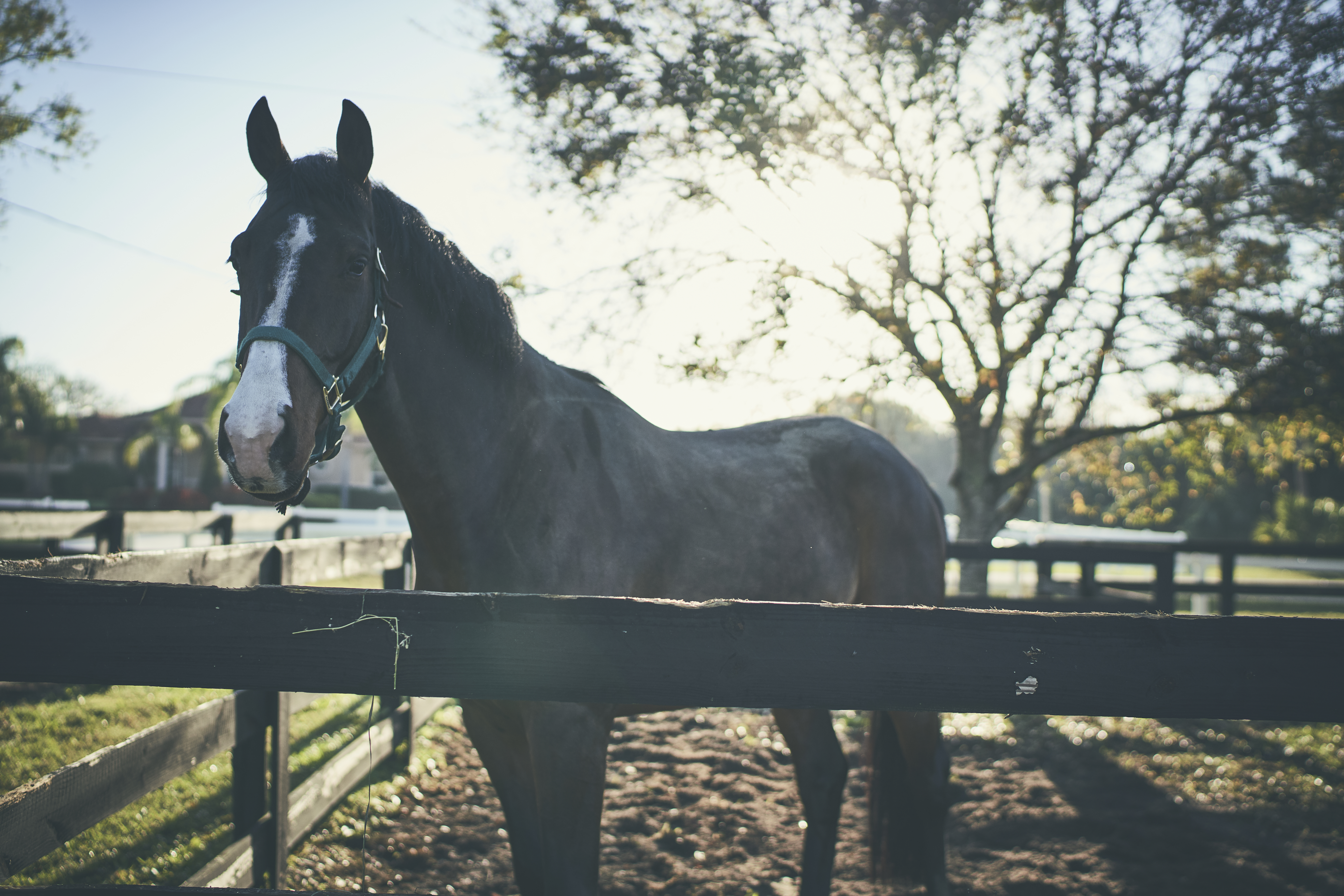
- Current accepted PPID prevalence is 21% of horses over the age of 15.1
- Although rare, horses as young as five years old have been diagnosed with PPID.
- PPID affects male and female horses, and has been identified in most breeds of horses and ponies.
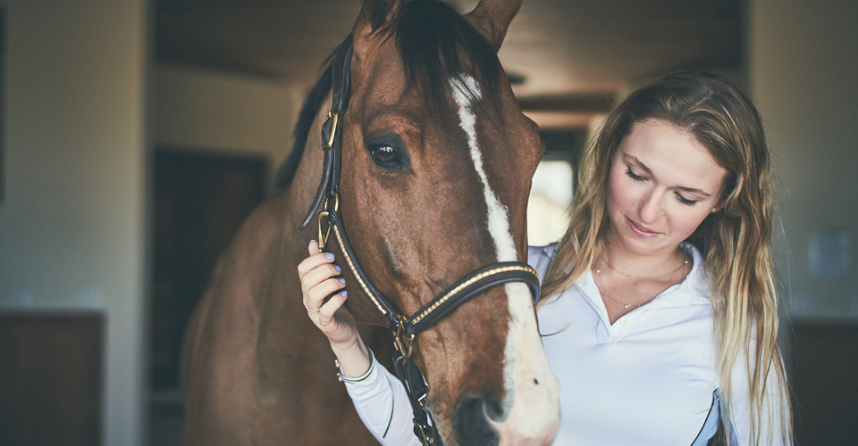
EARLIER THE DIAGNOSIS, THE BETTER
PPID is a chronic, degenerative disease. And while there are management options available, recognizing the clinical signs and early diagnosis is the first step leading to the proper management of a horse with PPID.
EARLY
A change in personality and/or a lack of energy.
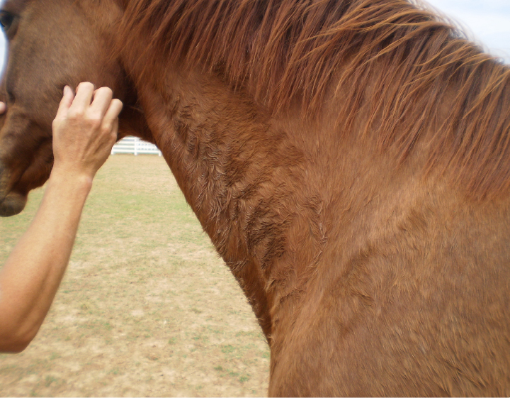
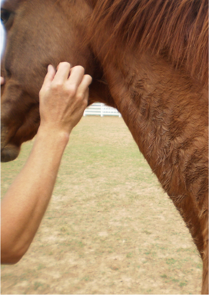
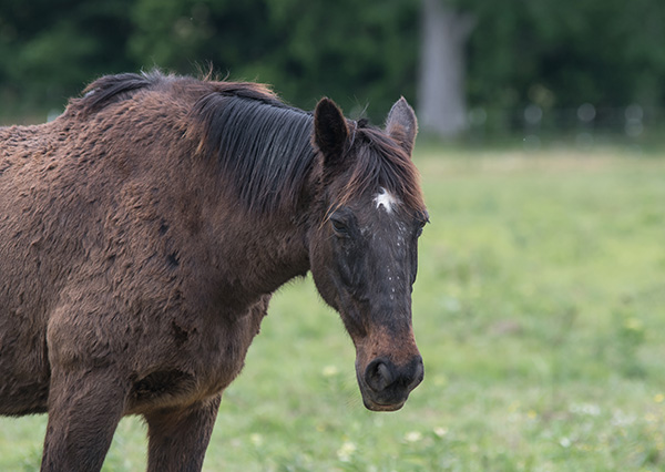
Decreased or delayed shedding in specific areas (regions) of the horse’s body.
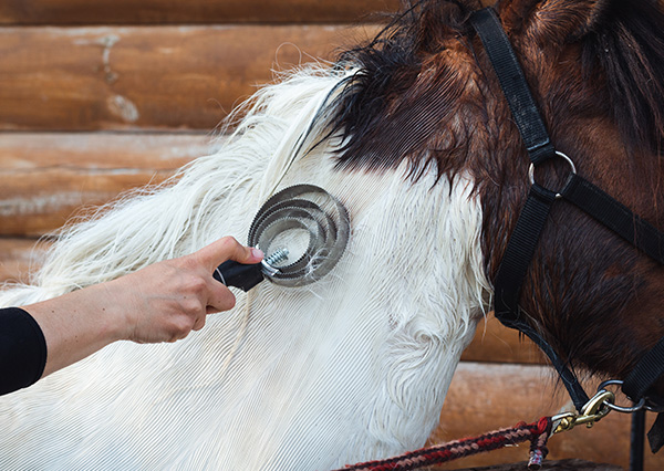
Delayed shedding of the winter coat may occur in some areas, and the summer coat may look different in these same areas.

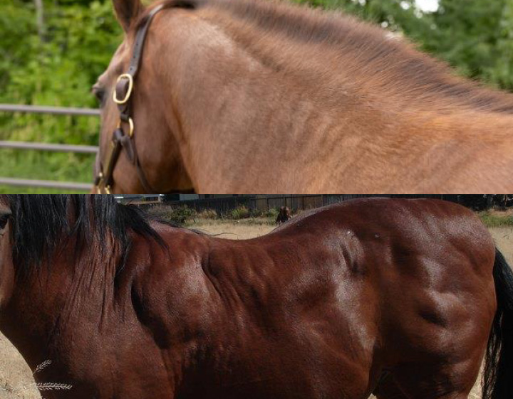
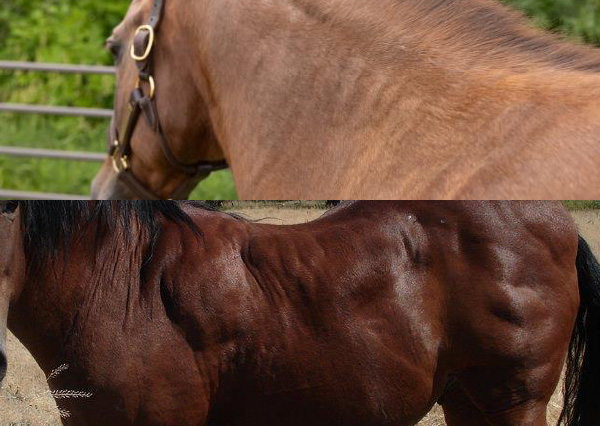
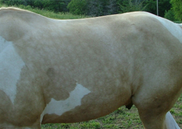
A gradual loss in muscle mass.
Increased or decreased sweating may occur.
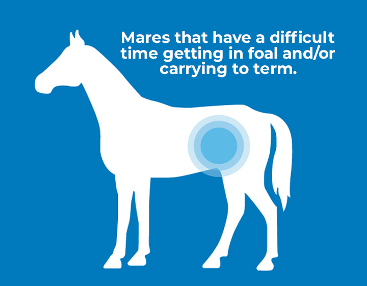
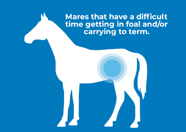
Reproductive cycles may be abnormal or absent, potentially leading to infertility.
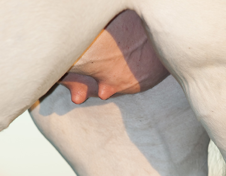
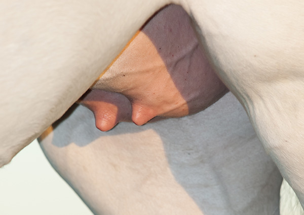
Fat deposits may appear along the crest of the neck and tail head.
Inflammation of the laminae of the foot and increased tenderness.


ADVANCED

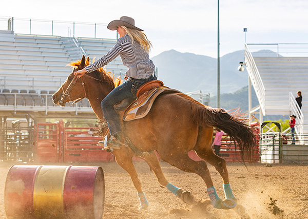
Sluggishness or decreased activity.


Decreased ability in physical exercise.
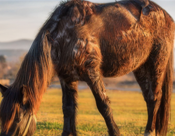
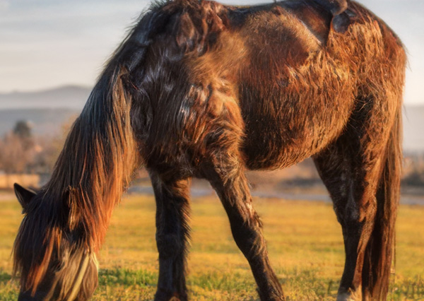
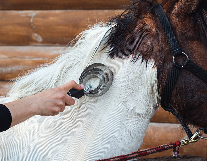
Excessive hair growth over all of the body.
Loss of seasonal shedding compared to herd mates or past years.

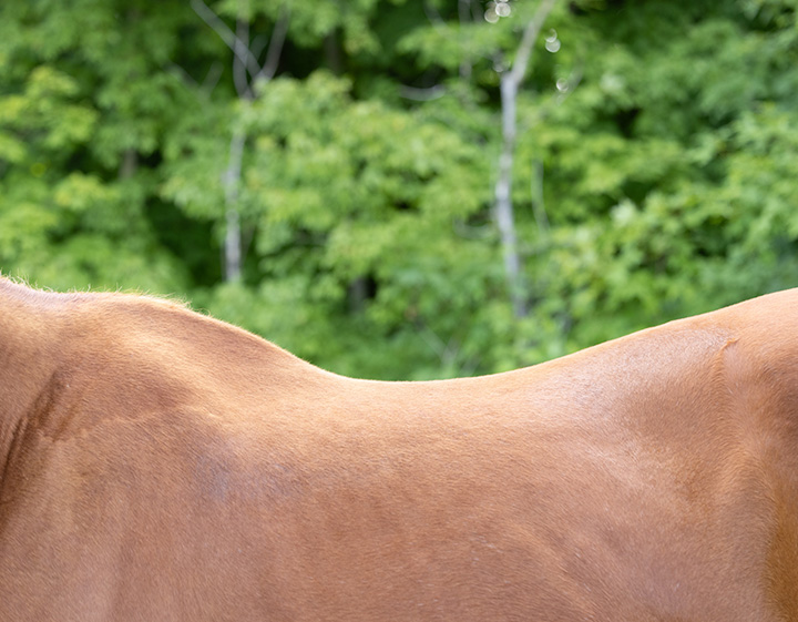
Prominent withers, sunken shoulders and/or prominent spine.

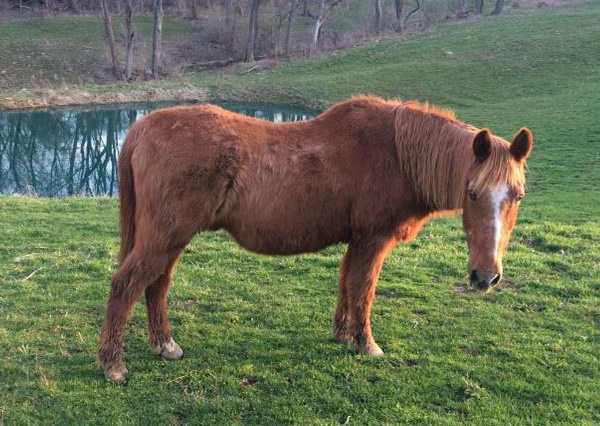
The horse's belly may have a "rounded" hay belly appearance.
Increased or decreased sweating may occur.
Increased urination and/or thirst.
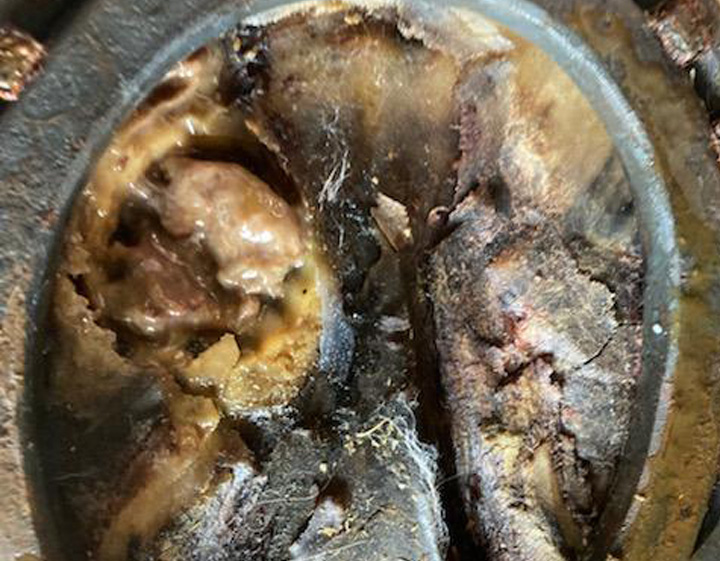

A few examples are recurring secondary skin infections, hoof abscesses, conjunctivitis and sinusitis.
Reproductive cycles may be abnormal or absent, potentially leading to infertility.
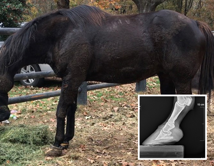
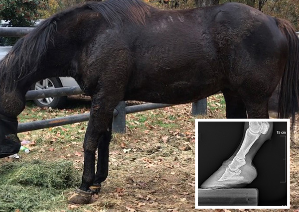
Tendon laxity may be observed where the fetlock "drops" toward the ground when bearing weight.
Inflammation of the laminae of the foot and increased tenderness.
Fat deposits may appear along the crest of the neck, the tail head and above the eyes.
REFERENCE
1 McGowan TW, Hodgson DR, McGowan CM. The prevalence of equine Cushing's syndrome in aged horses. In: Proceedings from the 25th American College of Veterinary Internal Medicine Forum; June 6-9, 2007; Seattle, WA. Abstract 603.
2 Grubbs ST, Neal DL and TJ Keefe. Epidemiological characteristics of horses at initial diagnosis. J Vet Intern Med 2015;29:123